For 40 years, we’ve helped scientists harness the full potential of biology with next-generation technology. Here we share the latest in automated, end-to-end solutions that span research disciplines to advance scientific discovery and improve the quality of human life worldwide.
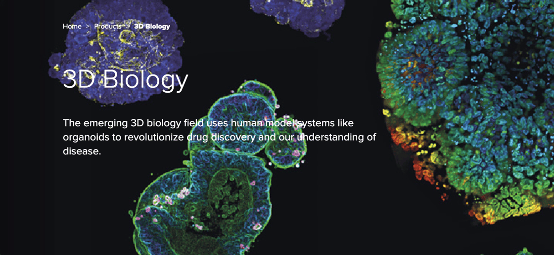
Unlock the full potential of 3D biology
The emerging 3D biology field uses human model systems like organoids to revolutionize drug discovery and our understanding of disease
Molecular Devices has been a life sciences industry leader for 40 years. We believe in the revolutionary promise of 3D cell models to advance next generation drug discovery. Whether you’re making the transition from 2D to 3D cell culture for the first time, scaling your organoid development program, or integrating a fully-automated screening workflow, we’re here to help.
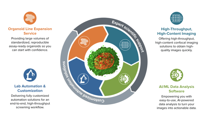
Custom Organoid Expansion Service- Provision of batches of standardized assay-ready organoids
Automated Bioprinting HCS Solution- Scale in-house organoid and 3D tissue development with an automated bio printer, high-content imager, and AI-powered image analysis software
Lab Automation & Customization - Streamline end-to-end workflows with next-gen 3D biology technologies that are customized into a fully-integrated, automated setup for any lab and supported by machine learning-enabled data analysis solutions
Growing organoids manually can present significant challenges for researchers that delay experiments and increase costs. Most common is an inability to produce sufficient numbers of organoids that are homogeneous in quality and size. Batch-to-batch consistency is virtually impossible without the standardized, repeatable processes employed in a regulated, industrial environment.
Molecular Devices has global leadership in the scale-up and industrial manufacturing of human-derived 3D organoids. Facilitated by our unique, patent-pending bioprocess, our semi-automated procedure uses controlled, monitored conditions to produce large numbers of standardized PDOs:
Ready to automate your lab for 3D models, but not sure where to start?
Bundle your workflow solution instruments and save!
There is a growing need for more biologically relevant models in drug discovery and disease modeling which has led to increased interest in three-dimensional models for assay development as they better represent the complexity of human tissue. However, reliability and scalability hurdles pose significant challenges to increasing adoption.
If your lab is struggling with issues such as workflow throughput, upstream sample prep, model reproducibility, and protocol standardization, we can help.
NEW!Research-ready organoids: Quickly scale up organoid expansion with solutions for the production of reproducible, assay-ready organoids
NEW! BAB400:Multifunctional robot automates organoid seeding in Matrigel domes, feeding, monitoring, incubation, and screening
The Role of CRISPR in Scientific Breakthroughs in Microbiome Engineering [Podcast]
Join leading experts Dr Jakob Haaber, Vice President & Head of Delivery Technologies, SNIPR Biome and Dr Richard Fox, Co-Founder, CEO and CTO of Infinome Biosciences, as they discuss the wide range of uses for CRISPR in microbiome engineering. Our accompanying blog will help summarize how to overcome the bottleneck of human microbiome research including for therapeutics and biomanufacturing:
How CRISPR-Cas9 helps explore the role of human microbiome in diseases
Advantages of CRISPR and recent developments
Acceleration in the manufacturing of CRISPR technologies
We caught up with Laura Dranschak, Director of Commercial Operations & Custom Solutions, to discuss all things SLAS 2023. See what all the buzz was about on the show floor, what the automation industry holds for labs of the future, and download our latest automation research poster on 3D complex assays:
Automating patient-derived colorectal cancer organoids for high-throughput screening
Automation of 3D bioprinting assays for high-content imaging and assessment of compound effects
Leveraging Automated Workflows to Enable Complex Organoid Assays
Novel analysis of neural outgrowth in 3D human brain micro-tissues
Optimizing Animal free CloneDetect assay with Protein G or A to maximize the sensitivity of real-time detection of human IgG antibody production for therapeutic protein engineering and cell line development using ClonePix System
Single-cell dispensing and screening of cell lines for monoclonality verification using the impedance-based single-cell dispenser and high-throughput fluorescence-based imager
How to implement cell line development and microbiology workflows
In case you missed it, or would like to view it again, our recent webinar on Colony Picking and Automation is now available to view on-demand.
Listen in as Janet Graystone, EU Sales Manager Biopharma, and Carola Mancini, Ph.D, EU Field Application Scientist Biopharma, discuss microbial colony picking and workflows, as well as cell line development processes in mammalian cell lines. Solutions to common challenges are presented, along with colony picking applications and case studies.
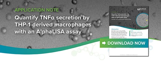
Quantify TNFa secretion by THP-1-derived macrophages with an AlphaLISA assay
In this application note, effects of several anti-inflammatory compounds on TNFα secretion by THP-1-derived macrophages were measured using the AlphaLISA High Performance (HP) Human Tumor Necrosis Factor alpha (TNFα) Detection Kit.
Using the SpectraMax i3x Multi-Mode Microplate Reader and this homogeneous assay, picogram levels of TNFα were detected in culture medium removed from THP-1 cells activated with PMA and LPS, and further treated with the compounds SB202190, stattic, and resveratrol.
Structural organization and functional analysis of compound response in 3D human iPSC-derived cardiac tri-culture microtissues
Recent publications show that tri-cellular co-culture microtissues of cardiomyocytes, endothelial cells, and cardiac fibroblasts that are all derived from human induced pluripotent stem cells (iPSCs) enhance the maturation and functional activity of cells compared to 2D cardiomyocytes and thus more closely mimics actual heart physiology.
In this application note, we developed methods for the formation of 3D spheroids derived from human induced pluripotent stem cells (iPSCs). Using both high-content imaging and fast kinetic fluorescence imaging, we measured the impact of various compounds on the beating rate and pattern of cardiac spheroids as monitored by changes in intracellular calcium levels with calcium-sensitive dyes.
In a recent study, we successfully knocked down the p53 protein in Human Embryonic Kidney cells (HEK-293) using CRISPR technology. The engineered cell line exhibited increased resistance to apoptosis-inducing compounds compared to control cells.
Our automated workflow played a crucial role in generating multiple engineered cell lines with specific mutations, opening doors to the development of cell-based disease models.
To understand the detailed methods and results of our study, please refer to the downloadable workflow poster, which provides comprehensive insights into stable transfection, monoclonality screening, and endpoint assays using our integrated system.
Please be advised that this email may contain confidential information. If you are not the intended recipient, please notify us by email by replying to the sender and delete this message. The sender disclaims that the content of this email constitutes an offer to enter into, or the acceptance of, any agreement; provided that the foregoing does not invalidate the binding effect of any digital or other electronic reproduction of a manual signature that is included in any attachment.