An intuitive and powerful platform for phenotypic profiling


Core Life Analytics' StratoMineR™ software helps biologists analyze the complex data derived from high-content image analysis. A powerful, intuitive workflow allows users to port data analyzed in IN Carta® Image Analysis Software directly into StratoMineR where it is used to generate rich, interactive visualizations using advanced data mining methods.

  • Built for biologists

    Built for biologists

    Intuitive analytics workflow. No coding required.

  • Phenotypic analysis

    Phenotypic analysis

    Use all of your high-content data to discover novel phenotypes.

  • Artificial Intelligence

    Artificial Intelligence

    Build your own AI models to discover the drugs of tomorrow

HC StratoMineR Introduction

HC StratoMineR Introduction

Features

  • Flexible data upload

    Flexible data upload

    Numerical data can be uploaded directly from IN Carta Image Analysis Software.

  • Artificial Intelligence

    Artificial Intelligence

    Machine learning models can be applied to leverage AI technology in data analytics.

  • Phenotypic characterization

    Phenotypic characterization

    Clustering and visualization of cellular phenotypes.

  • Quality Control

    Quality Control

    Use built-in data visualization tools for quick assessment of data quality and remove outliers.

  • Reduced data complexity

    Reduced data complexity

    Data reduction decreases complexity and increases focus on key biological interactions.

  •  Collaboration

    Collaboration

    Utilize project management tools for collaboration.

Import datasets from IN Carta for deeper insights into your data

IN Carta Image Analysis Software provides robust, quantitative results from complex biological images and datasets utilizing advanced AI technology. Directly import this data into StratoMineR, an intuitive web-based platform which guides users through a typical workflow in analysis of high-content, multi-parametric data.

StratoMineR intuitive workflow


Cluster analysis

StratoMineR Cluster analysis

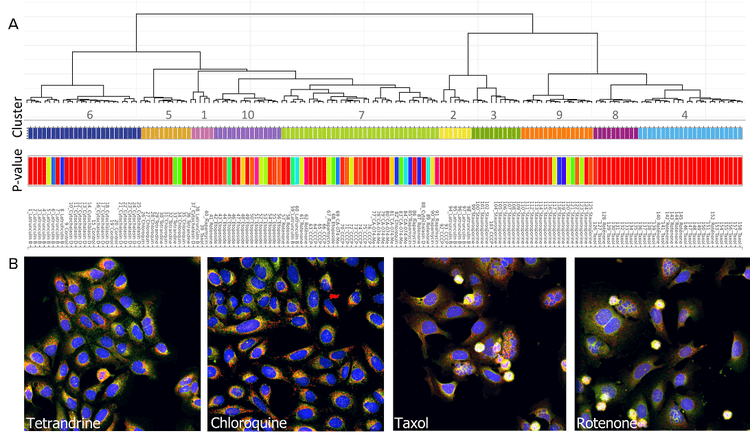
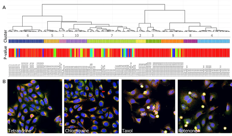
Cluster analysis: A) A dendrogram representing hierarchical relationships is shown. B) Examples of compound-treated cells belonging to the some of the clusters are shown. Cluster 5 consists of tetrandrine- and chloroquine-treated cells. Cluster 4 consists of rotenone- and paclitaxel-treated cells.

Scatter plot

Scatter plot

Example of a scatter plot using HC StratoMineR for visualization and exploration.

ImageXpress® Confocal HT.ai High-Content Imaging System

Powerful multi-laser light sources, a deep tissue penetrating confocal disk module, water immersion objectives and modern machine learning analysis software

New TagImagexpress confocal
  • Ideal for highly complex cell-based and 3D assays
  • Seven-channel high-intensity lasers generating brighter images with higher signal-to-background ratio
  • Spinning confocal disk technology for deeper tissue penetration, resulting in sharper images with improved resolution
  • Water immersion objectives offering up to quadruple the signal at lower exposure times for greater sensitivity and image clarity
  • View product

IN Carta Image Analysis Software

Modern machine learning, high-content image analysis software

CellReporterXpress software for ImageXpress Pico

Powerful analytics combined with an intuitive user interface simplify workflows for image analysis and phenotypic profiling. Advanced features provide the functionality you need to analyze data in 2D, 3D, and 4D - at scale - and deliver real-time insights without the need for complex pre- or post- processing operations. Improve specificity of your image analysis workflows by utilizing the SINAP deep-learning module and see for yourself that Segmentation Is Not A Problem. Put machine learning to work for you and perform complex phenotypic analysis within a user-friendly Phenoglyphs module.

Latest Resources

13

Applications of StratoMineR

  • Cell Painting

    Cell Painting

    Cell Painting is a high-content, multiplexed image-based assay used for cytological profiling. In a Cell Painting assay, up to six fluorescent dyes are used to label different components of the cell including the nucleus, the endoplasmic reticulum, mitochondria, cytoskeleton, the Golgi apparatus, and RNA. The goal is to “paint” as much of the cell as possible to capture a representative image of the whole cell.

    Learn More 

    Cellular Imaging & Analysis

    Cell Imaging & Analysis

    Researchers have several options in methods for imaging cells, from phase-contrast microscopy that shows intact cells to fluorescent imaging of single molecules or organelles. Cellular analysis is performed to evaluate and measure the current state of cells, such as cell integrity, toxicity, and viability and various other research applications. An integral part of cellular analysis is data collection, analysis, and export into a meaningful and useful format.

    Learn more 

  • Stem Cell Research

    Stem Cell Research

    Pluripotent stem cells can be used for studies in developmental biology or differentiated as a source for organ-specific cells and used for live or fixed cell-based assays on slides or in multi-well plates. The ImageXpress system has utility in all parts of the stem cell researcher’s workflow, from tracking differentiation, to quality control, to measuring functionality of specific cell types.

    Learn more 

    Toxicology

    Toxicology

    Toxicology is the study of adverse effects of natural or man-made chemicals on living organism. It is a growing concern in our world today as we are exposed to more and more chemicals, both in our environment and in the products we use.

    Read More 

Resources of StratoMineR

Presentations
Videos & Webinars
3D Imaging Seminar Series

3D Imaging Seminar Series

High-Content Phenotypic Screening

High-Content Phenotypic Screening

Emerging Organoid Models

Emerging Organoid Models: Translating Basic Research to Drug Development and Regenerative Medicine