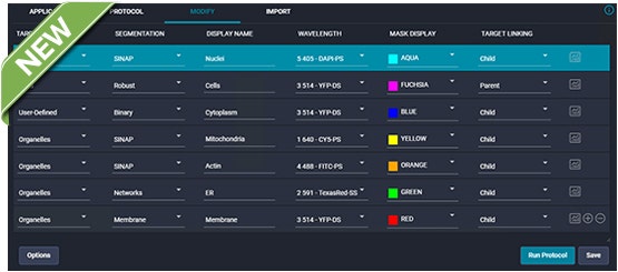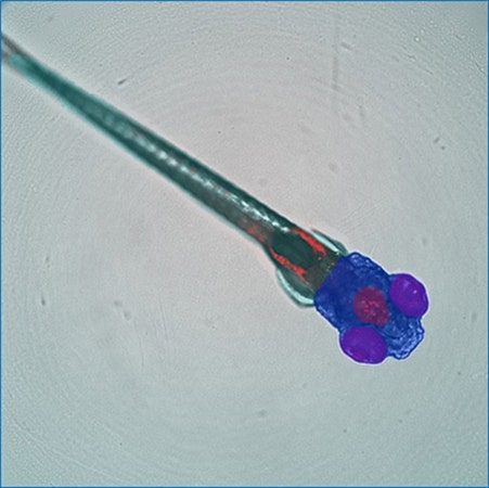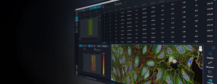
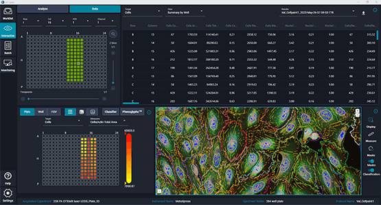
IN Carta® Image Analysis Software solves complex image analysis problems utilizing advanced Artificial Intelligence (AI) transforming images into results, which can be interpreted with ease. User-friendly workflows help you get answers faster from 2D, 3D, and 4D experiments. With the integration of our Custom Module Editor application, you can define highly customized image analysis protocols allowing you to obtain robust results—even for complex assays—then quickly visualize, review, and interact with the analysis results. Let IN Carta software do the heavy lifting so you can focus on your research.
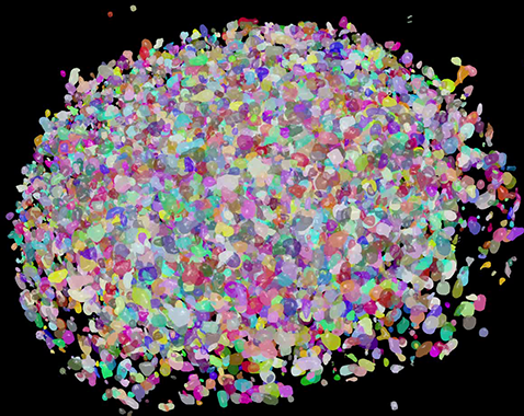
SINAP is a module that uses deep learning algorithms to improve accuracy and reliability of high-content screening assays at the first step in the analysis pipeline—segmentation. It provides better object detection than traditional image analysis methods. Deep learning models can be easily tailored within a user-friendly tool, so that any novel biological objects can be segmented efficiently. Quantitative information extracted from segmented objects is more accurate, so errors are not propagated down the analysis pipeline.
With SINAP, Segmentation Is Not A Problem!
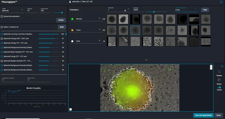
IN Carta® Phenoglyphs™ Software Module uses a unique combination of unsupervised and supervised machine learning to quantify phenotypical changes. Using many hundreds of cellular features that can be analyzed simultaneously, a comprehensive phenotypic profile is created and can be applied throughout an entire screening workflow. This multivariate approach to classification provides accurate characterization of object populations allowing users to resolve subtle phenotypic changes induced by drug treatment or genetic modification. It can be utilized across many biological targets including organoids, cells, spheroids, and more.
Powerful and intuitive workflows allow users to port high-content imaging data directly into StratoMineR where it is used to generate rich, interactive visualizations using advanced data mining methods. When used with IN Carta Image Analysis Software, it provides robust, quantitative results from complex biological images and datasets utilizing advanced AI technology. Use all of your high-content data to discover, characterize, and analyze Phenotypes.
Live cell imaging is the study of cellular structure and function in living cells via microscopy. It enables the visualization and quantitation of dynamic cellular processes in real time. Live cell imaging encompasses a broad range of topics and biological applications—whether it is performing long-term kinetic assays or fluorescently labeling live cells.
Toxicology is the study of adverse effects of natural or man-made chemicals on living organism. It is a growing concern in our world today as we are exposed to more and more chemicals, both in our environment and in the products we use.
Scientific Poster
Most potential oncology drugs fail at the later stages of the drug development pipeline and in clinical trials, despite having promising data for their efficacy in vitro. This high f…
Application Note
Patient derived organoids (PDOs) represent a promising tool to reduce pipeline attrition in drug discovery. These tumor organoids are multicellular mini replicas of the 3D tumor and…
Application Note
Many oncology drugs fail at the later stages of the drug development pipeline and in clinical trials, despite promising data for their efficacy in vitro. This high failure rate is…
Scientific Poster
3D organoids are increasingly popular in drug discovery and disease modeling as they better represent biologically relevant microenvironments, tissue architecture, and functionality. Ho…
Blog
Target discovery and drug development rely heavily on 2D cell and animal models to decipher efficacy and toxic effect of drug candidates. Yet, 90% of candidates fail to make it past…
Blog
Introduction – the problem. The average cost of bringing a new drug to the clinic is around $1 billion according to a study conducted by the London School of Economics in March 2020…
Blog
As we enter the era of sophisticated drug discovery with gene therapy and personalized medicine, we need to be prepared to study complex diseases, assess the therapeutic effect of…
Application Note
T-cell therapies are designed to help our immune system eliminate cancer cells. Those include CAR T-cells (Chimeric Antigen Receptor engineered T-cells), tumor infiltrating lymphocytes…
Brochure
The BioAssemblyBot® 400 is an intelligent robot used by life scientists to build 3D model systems with increased throughput and precision, alleviating major concerns mentioned above.…
Publications
Gone are the days of only measuring single parameters in cell-based experiments. Instead, researchers should widen their focus, namely with the help of innovations like the Cell Pain…
Scientific Poster
Most potential oncology drugs fail the drug development pipeline, despite having promising data for their efficacy in vitro. This further incentivize the need for identifying in vitr…
Scientific Poster
Three-dimensional (3D) cell models that represent various tissues are being successfully used in drug discovery and disease modeling to study complex biological effects and tissue ar…
eBook
Complex biological systems such as spheroids, organoids, and organ-on-a-chip are becoming more popular for disease modeling and drug screening as they better simulate organs and tiss…
Application Note
The human brain is highly complex which makes it challenging to study both in vitro and in-model organisms. Cultured neurons do not sufficiently recapitulate the three-dimensional (3D…
Application Note
The intestinal crypt system is an important part of the intestinal organoid where mature organoids have more complex and numerous crypt structures. Yet, due to their high turnover rate…
Blog
It was another great year at ISSCR 2022! Leaders from across the globe came together to discuss new technologies, share insights, and explore the newest breakthroughs in stem cell…
Publications
Today, nine out of ten drugs fail in clinical trials. Furthermore, it takes over a decade and an average cost of $2 billion to develop and approve each…
Scientific Poster
The culture of complex organoids such as the cerebral organoid is a rapidly developing technology that has immense potential in areas of developmental neurobiology and neurodegenerat…
Scientific Poster
3D cell models representing various tissues were successfully used for studying complex biological effects and tissue architecture, however, the complexity of 3D models remains a hur…
Blog
3D cell models are becoming increasingly popular for studying complex biological effects, tissue functionality, and diseases. Their ability to self-organize and mimic…
eBook
Three-dimensional (3D) organoid development is one of the most important advancements in drug discovery research to date.
Application Note
Triple negative breast cancer is a clinically aggressive tumor subtype, with high rates of metastasis, recurrence, and drug resistance.
Publications
Cell painting is a high-content profiling technology that uses up to six fluorescent dyes to visualize specific cellular components at the single-cell level. The assay essentially “p…
Brochure
Core Life Analytics’ StratoMineR™ software helps biologists analyze the complex data derived from high-content image analysis. A powerful, intuitive workflow allows users to port dat…
Application Note
Cell painting has become a popular screening method within the drug discovery community. This phenotypic screening platform leverages advances in high-content microscopy, high-…
Publications
Three-dimensional (3D) cell-based assays replace a host of tissue, cell, and animal test models but have a reputation for lacking time resolution. For e…
Scientific Poster
Multiparametric high-content screening approaches, such as the Cell Painting assay, are increasingly being used in many applications ranging from drug discovery programs to functiona…
Scientific Poster
Here we describe a workflow for automation of organoid culture. The automated method utilizes an integrated work-cell, consisting of several instruments providing automated cell cult…
Application Note
Label-free cell analysis provides a better alternative to using fluorescent dyes as it enables scientists to image live cells under near-native conditions – examining biological…
Blog
SLAS2022, the Society for Lab Automation and Screening conference offered another exciting year for learning about innovative laboratory technologies. Whether you attended in-person…
Application Note
Image-based phenotypic profiling approaches such as the widely used Cell Painting assay use high-content imaging along with multiparametric readouts to study biological, genetic, and…
Scientific Poster
3D cell culture as a model system is increasingly popular because it recapitulates the in vivo microenvironment better than 2D cell cultures. Organoids have the capacity for stable d…
Scientific Poster
Triple negative breast cancer is a clinically aggressive tumor subtype, with high rates of metastasis, recurrence, and drug resistance. Currently there are no clinically approved sma…
Scientific Poster
Image-based phenotypic profiling approaches, such as the widely-used Cell Painting assay, use high-content imaging along with multiparametric readouts to study biological, genetic, a…
Application Note
Complex 3D biological models such as organoids and patient-derived spheroids are gaining popularity in many biomedical research areas because they more closely recapitulate the in vivo…
Blog
From customer feedback to workflow improvements The path to understanding complex biological processes and diseases is paved with a lot of challenges. As the desired level of…
Blog
If you didn't get a chance to visit us at our poster sessions during ISSCR 2021, don’t fret. We've gathered all our sessions right here for you. The ISSCR Annual Meeting brought…
Application Note
We describe an automated integrated system that would allow automated monitoring, maintenance, and characterization of growth and differentiation of organoids and stem cells, as well…
Publications
Lab Manager speaks to Dan O’Connor, vice president, drug discovery, Molecular Devices, about the company’s Organoid Innovation Center in San Jose, CA. The center is 180 sq. ft., with…
Publications
Humans began culturing animal cells and tissues as early as the late 19th century, when Wilhelm Roux first showed that chick embryos could be cultured in saline solution for a few da…
Brochure
IN Carta™ Image Analysis Software solves complex image analysis problems utilizing advanced Artificial Intelligence (AI) turning images into data. Easy-to-use workflows help you get…
Blog
There have been significant advancements in microscopy and camera technology, as well as advancements in technologies for labeling molecules of interest over the past decade. These…
Publications
Molecular Devices, a provider of innovative life science technology, recently unveiled a brand new, first-of-its-kind Organoid Innovation Center. Situated at the company’s global hea…
Blog
Artificial intelligence (AI) is finding its way into many aspects of modern life, from autonomous vehicles to voice-powered personal assistants, and even the creation of art. But it…
Blog
Have you ever heard the old adage, “A picture is worth a thousand words?” When it comes to Cell Painting, this saying is especially true. Cell Painting is a high-content,…
Publications
It has been a busy beginning of the year for Danaher’s Life Sciences segment. In January, the business reported 2020 revenues of $10.6 billion, represen…
Blog
For over 40 years, Molecular Devices has been at the forefront of technological advances which have contributed to significant scientific breakthroughs. To kick off the new year, we…
Publications
Developments in informatics solutions and high-content imaging instruments, increased focus on cell-based research, and rising government support of research and development for drug…
Brochure
Molecular Devices is a leading provider of high-performance bioanalytical measurement solutions for life science research, pharmaceutical development, and biotherapeutic discovery. O…
Automating 3D Organoid Models and Assay Workflows
Using Differentiated iPSCs to Build Ready-to-use 3D Models
The search for answers: Using lab automation with patient-derived tumoroids to find more relevant therapies for clinically aggressive cancers
3D Imaging Seminar Series
New standard in organoid culture and image-based analyses
Automate your 3D biology high-throughput workflows
Leverage automated, end-to-end workflows to enable complex organoid assays
Level Up your 3D Cell Culture: From Research to High-Throughput
Enhance high-content 3D biology imaging with automated sample preparation
Enhancing 3D Disease Models: Automated, High-Throughput, Phenotypic Screening with Organ-on-a-Chip
High-Content Phenotypic Screening
Emerging Organoid Models: Translating Basic Research to Drug Development and Regenerative Medicine
Automating culture and high-content imaging of 3D organoids for in vitro assessment of compound effects
Deep Learning-Based Image Analysis for Label-Free Live Monitoring of iPSC and 3D Organoid Cultures
Monitoring organoid development in iPSC-derived 3D cerebral organoids
ISSCR 2021 Innovation Showcase: Automated Culture and High-Content Imaging of 3D Lung and Cardiac
Senescence occurs in response to a number of damaging stimuli to limit oncogenic transformation and cancer development. As no single, universal senescence marker has been discovered, the confident classification of senescence induction requires the parallel assessment of a series of hallmarks. Therefore, there is a growing need for “first-pass” tools of senescence identification to streamline experimental workflows and complement conventional markers.
The COVID-19 pandemic caused by the SARS-CoV-2 virus has led to a major public health burden and has resulted in millions of deaths worldwide. As effective treatments are limited, there is a significant requirement for high-throughput, low resource methods for the discovery of novel antivirals. The SARS-CoV-2 spike protein plays a key role in viral entry and has been identified as a therapeutic target.
Cathepsin L is a key host cysteine protease utilized by coronaviruses for cell entry and is a promising drug target for novel antivirals against SARS-CoV-2. The marine natural product gallinamide A and several synthetic analogues were identified as potent inhibitors of cathepsin L with IC50 values in the picomolar range. Lead molecules possessed selectivity over other cathepsins and alternative host proteases involved in viral entry. Gallinamide A directly interacted with cathepsin L in cells and, together with two lead analogues, potently inhibited SARS-CoV-2 infection in vitro, with EC50 values in the nanomolar range. Reduced antiviral activity was observed in cells overexpressing transmembrane protease, serine 2 (TMPRSS2); however, a synergistic improvement in antiviral activity was achieved when combined with a TMPRSS2 inhibitor. These data highlight the potential of cathepsin L as a COVID-19 drug target as well as the likely need to inhibit multiple routes of viral entry to achieve efficacy.
To analyze (1) the effect of immunoglobulin G (IgG) from patients with anti–myelin oligodendrocyte glycoprotein antibody (MOG-Ab)–associated disorder on the blood-brain barrier (BBB) endothelial cells and (2) the positivity of glucose-regulated protein 78 (GRP78) antibodies in MOG-Ab–associated disorders.
The development of a safe and effective vaccine is a key requirement to overcoming the COVID-19 pandemic. Recombinant proteins represent the most reliable and safe vaccine approach but generally require a suitable adjuvant for robust and durable immunity. We used the SARS-CoV-2 genomic sequence and in silico structural modelling to design a recombinant spike protein vaccine (Covax-19™).
Considerable concerns relating to the duration of protective immunity against severe acute respiratory syndrome-coronavirus-2 (SARS-CoV-2) exist, with evidence of antibody titers declining rapidly after infection and reports of reinfection. Here, we monitor the antibody responses against SARS-CoV-2 receptor-binding domain (RBD) for up to 6 months after infection. While antibody titers are maintained, ∼13% of the cohort’s neutralizing responses return to background.