
Bioneer use the ImageXpress Micro Confocal for high-throughput imaging of 3D disease models
COMPANY/UNIVERSITY
Bioneer A/S
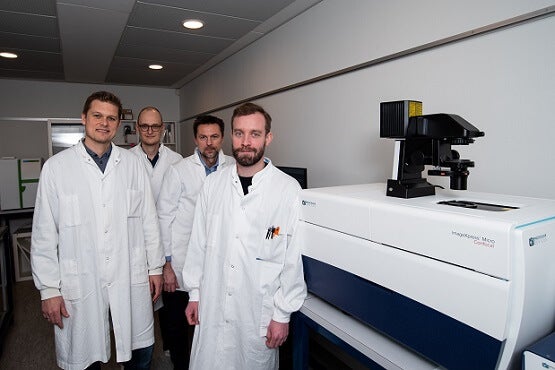
TEAM MEMBERS
Christian Clausen
Bjørn Holst
PRODUCTS USED
ImageXpress Micro Confocal High-Content Imaging System
The Challenge
Revolutionizing early drug discovery for immuno-oncology and neurodegenerative disease modelling: High throughput imaging of 3D models of disease
Bioneer A/S is a Danish research-based service provider, offering advanced cellular assays, molecular histology techniques and biomarker discovery, protein production and drug development services. Originally established in 1982, they are headquartered north of Copenhagen and have another two sites in Copenhagen and Lund, Sweden. Their mission is to increase the competitiveness of their global customers by facilitating their early, explorative development of new therapeutic solutions.
Christian Clausen, Director of Research and Development at Bioneer, says “We have state-of-the-art laboratory facilities and scientifically trained employees that strive to solve R&D projects from the very simple ones to highly complex projects, involving multiple disciplines across our organization. One brilliant example is 3D disease models, where a multi-disciplinary approach is required to provide customers with data from a complicated, more i n vivo-like setting”.
Bioneer has been developing cell models and cellular assays for companies for many years with accumulating proficiency. They have especially been focused on immune models, cancer models, neurodegenerative models and a range of barrier models (e.g., blood-brain barrier and intestinal models) as well as quantitative imaging. In recent years, it has become more and more clear to them that moving these models and technologies into a combination of 3D and high-throughput setups would give a range of new ways to solve early drug discovery projects for our customers.
Four years ago, the team initiated internal development programs to move their in-house models and cellular assays into 3D formats, in semi-high-throughput configurations for proof-of-concept studies. These efforts have been very successful, and the team are now at a stage where implementing some of the models into high-throughput 3D imaging platforms is the next step.
After evaluating different 3D high-throughput imaging systems, the team chose the ImageXpress Micro Confocal High-Content Imaging System to advance their immuno-oncology and neurodegenerative diseases studies.
The Solution
Christian Clausen says, “It is not only important to have the best cellular models for Bioneer, but also to have state-of-the-art imaging and analysis equipment to offer high-qualitative research support to our customers. We believe that the features of the ImageXpress Micro Confocal system fit our needs and will further potentiate our ability to assist small to midsize biotech companies without knowhow and/or equipment to do these analyses in-house, as well as large pharma companies who either need help with implementing the technology in-house or are looking for an external partner to handle these analyses in the drug discovery process”.
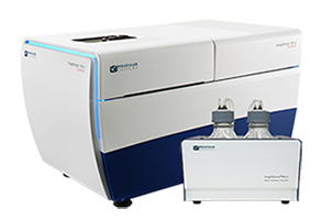
ImageXpress Micro Confocal High-Content Imaging System
Products Used
The ImageXpress Micro Confocal system is a high-content solution that can switch between widefield and confocal imaging of fixed and live cells. It can capture high quality images of whole organisms, thick tissues, 2D and 3D models, and cellular or intracellular events. The spinning disc confocal and sCMOS camera enable imaging of fast and rare events like cardiac cell beating and stem cell differentiation. With the MetaXpress software, the system enables many confocal imaging applications from 3D assay development to screening.
The Results
Immuno-oncology
The team at Bioneer have seen an increasing demand and interest for models that are not just single-layered cells (2D), but where the cells have had the possibility to form structures that more closely resemble the tumor microenvironment seen in vivo. This also includes self-organization with other cell types like stroma cells. The cutting edge of the immuno-oncology field is to study the function of the immune cells and their ability to recognize and kill cancer cells. They are now working on establishing 3D cancer spheroid models, combining their oncology competences with their immune modeling platform. Using this model, they will be able to analyze the infiltration of immune cells (T cells and NK cells) into different tumors. Thanks to the implementation of the ImageXpress Micro Confocal, they aim at establishing high-throughput assays for screening various small molecules and checkpoint inhibitors for their effects on the 3D immune-cancer spheroids. They will also analyze the mode-of-action of immune cells to kill or migrate into the cancer spheroids, as well as the influence of different candidate immuno-oncology drugs.
Neurodegenerative disease
Using human induced pluripotent stem (iPS) cells, the Bioneer team have built models for several neurodegenerative diseases like Alzheimer’s, Parkinson’s and Frontotemporal dementia. They are already well on their way to developing different assays reflecting disease-specific phenotypes in these models. Such phenotypic assays could, for example, be characterization of protein aggregates, mis-localization of proteins, autophagy, mitophagy and neurite outgrowth. As a proof-of-principle system for neurite outgrowth, they have developed an iPS cell line where they can induce expression of the NGN2 protein, which leads to accelerated differentiation of the cells to neurons with neurite outgrowth already occurring after 2-3 days. The team are now implementing this on the ImageXpress Micro Confocal to monitor neurite outgrowth in more advanced setups. They will also use the well-described phenotypes – like altered function of the lysosomes and cellular autophagy – to set up high-throughput 3D imaging assays using the ImageXpress Micro Confocal to analyze pharmaceutical compounds affecting these processes.
Drug discovery now relies mainly on high-throughput assays with a relevant window for measuring the effects of candidate drugs. 3D cell models, in general, are expected to revolutionize the output from early drug discovery, potentially resulting in a more qualified early selection of lead candidates and, subsequently, improved R&D productivity. Bioneer has an optimal standpoint in this field, in terms of competencies and technologies, and will be able to significantly impact early drug discovery approaches in both biotech and large pharma companies. Especially within immuno-oncology and neurodegenerative disease modeling, Bioneer will be an optimal partner to work with.

Bioprinted 3D culture of inducible neurons analyzed on the ImageXpress Micro Confocal High-Content Imaging System. Green = GFP. Red = β-Tubulin. Blue = Hoechst. Image courtesy of Bioneer A/S.