
Application Note
High-content cell profiling using a next-generation imaging platform with LED illumination
- Accelerates high-content imaging workflows with faster plate acquisition enabled by optimized stage motion, adaptive autofocus, and efficient widefield LED illumination while maintaining high signal-to-noise image quality.
- Enables reliable quantification of complex cellular phenotypes through accurate segmentation of nuclei, cells, and subcellular structures using AI-powered analysis tools within IN Carta® Image Analysis Software.
- Supports flexible multichannel imaging across diverse assays with stable LED illumination that enables rapid wavelength switching while reducing phototoxicity and photobleaching in fixed and live-cell experiments.
Zhisong Tong | Research Scientist | Molecular Devices
Angeline Lim | Product Manager, Imaging | Molecular Devices
Introduction
High-content imaging has become an essential tool across a wide array of biological research, enabling researchers to visualize, quantify, and analyze complex cellular responses across multiple spatial and functional dimensions. As biological questions increasingly require multiparametric, live cell, and high resolution data, imaging systems must provide reliable quantitative data, while remaining accessible and easy to use across diverse experimental workflows and user expertise.
The ImageXpress® HCS.ai High Content Screening System was developed to support these wide-ranging research needs through a fully redesigned hardware and software platform optimized for performance and usability.Improvements in stage motion and adaptive autofocus enable fast, precise image acquisition, while an optimized optical path and high–quantum efficiency camera deliver bright, high contrast images with reduced background and shorter exposure times.
Widefield imaging with LED illumination provides a practical and versatile approach for many applications in multi-user academic labs, particularly for 2D samples, live cells, and multiparametric experiments. LED illumination offers stable and reproducible light output, rapid wavelength switching without warm up, with reduced phototoxicity and photobleaching. These characteristics make the ImageXpress HCS.ai Widefield System (Figure 1) well suited for flexible multichannel widefield imaging to accommodate a broad range of research applications.
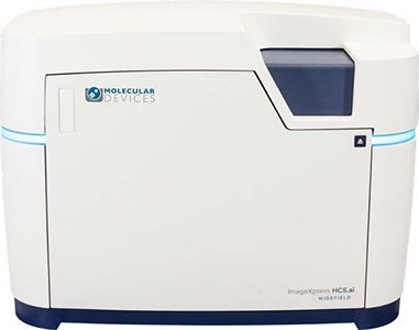
Figure 1. ImageXpress HCS.ai Widefield System
In this application note, we demonstrate the performance of the ImageXpress HCS.ai widefield system using a G protein–coupled receptor (GPCR) β arrestin translocation assay as a representative example of quantitative cellular imaging. The study highlights a streamlined, end to end workflow—from image acquisition to data analysis—and illustrates how the system enables rapid image collection while maintaining the image quality and measurement consistency required for reproducible biological research.
Methods
Cell culture and staining
U2OS cells expressing GFP tagged β arrestin were plated in a 384 well flat bottom plate (Greiner) and maintained in Dulbecco’s Modified Eagle Medium (DMEM; Thermo Fisher Scientific) for 24 hours. The cells were then exposed to the GPCR agonist isoproterenol (SelleckChem) for one hour. Following treatment, cells were stained with MitoTracker Deep Red (Thermo Fisher Scientific) and Hoechst (Thermo Fisher Scientific), and subsequently fixed with 4% paraformaldehyde (PFA; Thermo Fisher Scientific). After fixation, the solution was removed, and the wells were washed twice with PBS before imaging.
Image acquisition and analysis
Images were acquired on the ImageXpress HCS.ai High-Content Screening System using 20X magnification, Widefield LED illumination in DAPI, FTIC and TL channels. A single-z plane of each channel was acquired. The segmentation of nuclei and G-protein coupled receptor (GPCR) puncta was done with IN Carta® Image Analysis Software. The nuclei were segmented with built-in Nuclei Robust tool, the GPCR were segmented with built-in Organelles Robust Puncta tool and Cells were segmented with built-in SINAP Cellpose model with Diameter parameter set as 50 µm and Reference Wavelength parameter set as DAPI (Figure 5A). Puncta counts on a per-cell basis were quantified to construct the dose-response curve.
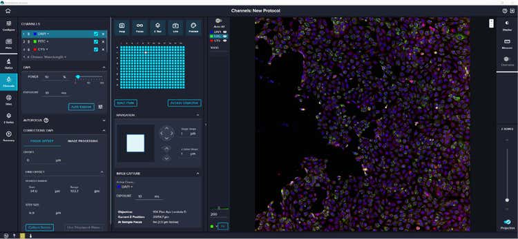
Figure 2. Redesigned image acquisition software (MetaXpress® Acquire High-Content Image Acquisition Software) offers an intuitive, user-friendly environment that is quick for novices to master while remaining flexible and customizable for advanced users.
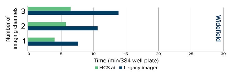
Figure 3. Average acquisition speeds of three runs for each acquisition setup shown (2 instruments, 20X). The acquisition was performed with widefield. Only 2D imaging was performed.
Results
Up to 100% faster acquisition speed with widefield
The ImageXpress HCS.ai widefield system is optimized for both speed and assay flexibility. To evaluate imaging performance, we compared image acquisition times across several configurations and benchmarked the system against legacy imagers. Plate acquisition time with widefield was, on average, 50% shorter, representing a 100% improvement in speed (Figure 3). Using identical exposure times across systems, the ImageXpress HCS.ai widefield system produced pixel intensities that were at least twice as bright and delivered at least a 2 fold improvement in signal to noise ratio.
Cell profiling performance with widefield and LED illumination
G protein coupled receptors (GPCRs) are the largest class of pharmaceutical targets and are commonly used in cell based screening assays. We used a genetically engineered cell line that allows for visualization of β arrestin translocation. When GPCRs are activated, cytoplasmic β arrestin translocates to membrane pits and endocytic vesicles. In this study, U2OS cells expressing GFP tagged β arrestin were treated with isoproterenol, a GPCR agonist, enabling high content imaging and analysis of internalized GPCR vesicles (Figure 4A).
Accurate cell segmentation is essential for reliable counting of vesicle puncta and quantifying dose response effects. IN Carta Image Analysis Software incorporates Cellpose 3.0 to deliver precise cell segmentation, displaying masks in randomized colors to clearly differentiate neighboring cells (Figure 4C). Cellpose is a deep learning– based algorithm capable of segmenting a wide range of cell images without retraining. Its robustness enables it to handle variations in noise, blur, contrast, imaging channels, and object sizes. For GPCR vesicles segmentation, the Organelles Robust Puncta tool was implemented in IN Carta Image Analysis Software (Figure 4E).
Enumerated GFP puncta were used to calculate the z-prime score (0.76 and 0.77) and EC50 for the assay (3.13 nM and 3.40 nM) (Figure 5B and 5C).
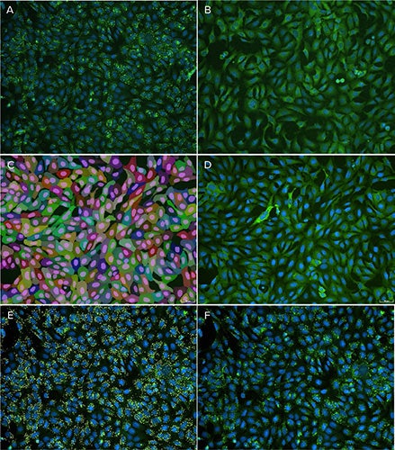
Figure 4. A. Cells with activated GPCR (treated with isoproterenol), note the presence of β arrestin puncta (green) in the treated sample; B. Negative control; C. Cell masks in random colors, generated using Cellpose model in IN Carta software with the corresponding raw image in D; E. Puncta masks in yellow color generated using Organelles Robust Puncta tool in IN Carta software with corresponding raw image in F.
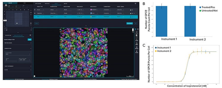
Figure 5. A. Image analysis protocol: Nuclei were segmented with Nuclei Robust tool on DAPI channel; Vesicles were segmented with Organelles Robust Puncta tool on FITC channel; Cells were segmented with Cellpose 3.0 on FITC channel with reference to DAPI channel. B. Average number of puncta per cell from two ImageXpress HCS.ai systems with LED and widefield illumination. The z’ scores are 0.76 and 0.77, respectively. C. The dose-dependent increase in the number of puncta per cell, showing activation of G-PCR pathway in response to isoproterenol. The EC50 values, determined using a four-parameter logistic regression model, were 3.13 nM for instrument 1 and 3.40 nM for instrument 2.
Conclusion
The ImageXpress HCS.ai High Content Screening System delivers a powerful and flexible platform for quantitative cell biology research. Its redesigned hardware, adaptive autofocus, and optimized LED based widefield illumination enable rapid image acquisition, high signal to noise ratios, and reduced phototoxicity and photobleaching, thus supporting a wide range of fixed and live cell imaging experiments.
Using a GPCR β arrestin translocation assay, the system demonstrated robust and reproducible performance, with consistent quantitative measurements across instruments.
Advanced analysis tools within IN Carta Image Analysis Software enabled accurate cell and subcellular segmentation, facilitating reliable extraction of biologically meaningful features with minimal manual intervention.
Together, these capabilities support a seamless workflow from image acquisition through data analysis, making the ImageXpress HCS.ai widefield system well suited for academic multi-user laboratories. By combining ease of use with high quality quantitative imaging, the system empowers researchers to focus on biological discovery while generating reproducible, ready data.