
Application Note
Automated expansion and monitoring of patient-derived CRC organoids using the CellXpress.ai system
- Remove the manual burden of routine colorectal cancer organoid (CRC) organoid maintenance by consolidating seeding, feeding, imaging, and passaging into a walk away workflow.
- Substantially reduce hands on time and generating assay-ready organoids for downstream applications.
- Automate CRC cultures to ensure consistent growth and reduced variability.
- Characterize and passage long-term healthy CRC organoid cultures using AI-driven analysis, deep learning segmentation, and classification.
Krishna Macha, Oksana Sirenko, Nikki Carter | Molecular Devices, LLC
Introduction
Three-dimensional (3D) patient-derived CRC organoids have emerged as powerful preclinical models that preserve tumor architecture, genetic heterogeneity, and drug-response phenotypes more accurately than traditional 2D cultures. Their biological relevance makes them valuable for disease modeling, compound screening, and precision oncology; however, routine culture of CRC organoids remains technically demanding. Manual handling of Matrigel domes, media exchanges, and passaging introduces variability, restricts scale, and limits the ability to generate large, consistent batches of assay-ready organoids for systematic testing. These challenges significantly hinder the reproducibility and throughput required for translational and drug discovery workflows.
To address limitations in scalability and standardization, automated culture platforms have become increasingly important. The CellXpress.ai® Automated Cell Culture System integrates liquid handling, environmental control, transmitted light imaging, and AI-enabled analysis to deliver a fully automated, closed-loop system for organoid maintenance and expansion. The platform uses IN Carta® Image Analysis Software’s deep learning segmentation module and Phenoglyphs™ based classification to objectively assess organoid morphology, maturity, density, and other complex phenotypic features. These quantitative metrics support rule-based decisions for feeding and passaging, reducing operator-dependent variability and enabling stable long-term culture. Such automation is particularly critical for CRC organoids, which require precise dome handling and careful timing of fragmentation to maintain epithelial structure and viability over extended periods.
The system not only increases throughput, but also improves consistency and reproducibility, making it easier to generate large cohorts of assay-ready organoids for high-frequency screening and downstream applications.
In this study, we implemented a complete workflow using the CellXpress.ai system for patient-derived CRC organoid expansion, maintenance, and assay preparation. Beginning with a vial of 3D Ready™ CRC organoids, we seeded multiplate cultures, performed scheduled and AI-guided media exchanges and passaging, and scaled cultures for more than four weeks. The automated workflow expanded one vial of starting organoids, which were then harvested and reseeded into 96-well plates for downstream applications. This automated workflow also minimizes routine manual handling, allowing longterm CRC organoid expansion to proceed with minimal oversight while greatly reducing the operational burden on researchers.
To assess organoid quality following automated expansion, we used confocal imaging to assess epithelial integrity and organization through phalloidin, ZO-1, and DAPI staining.
Overall, this work demonstrates that combining automation with AI-driven analysis provides a robust, reproducible, and scalable approach for CRC organoid expansion, enabling consistent production of assay-ready cultures for high-throughput screening and translational oncology studies.
Materials and methods
Organoid seeding, feeding, and monitoring – a vial of colorectal cancer organoids (ISO68, Molecular Devices) containing 100,000 organoids was thawed in a water bath and processed according to the provided protocol. The organoids were resuspended in 20% complete media (DMEM/F12 [Gibco, Cat. No. 11039] + 10% FBS, B27 [Invitrogen, Thermo Fisher Scientific], ROCK inhibitor [Tocris Bioscience], N-acetyl cysteine [Sigma-Aldrich]) and 80% GF-reduced Matrigel (Corning, Cat. No. 356231). The suspension was transferred to a 96-deep well reservoir for seeding into 24-well culture plates on the CellXpress.ai system. Organoids were seeded at a density of 800 organoids per well. During organoid culture, automated media exchanges were performed every 48 hours, and transmitted-light imaging was conducted every 24 hours (Figures 3–5). The imaging protocol utilized a 4x objective with plate bottom autofocusing and a 3 x 3 tiled field of view to generate high coverage transmitted light images. A 5 step Z series with 100 µm spacing provided reliable best focus projection across samples with variable thickness.
Organoids were analyzed using the IN Carta software, which employs deep learning segmentation within the Segmentation is Not A Problem (SINAP) module. A custom analysis protocol was created using the Flex-Protocol application to extract features across wells. The Phenoglyphs software module then performed machine learning (ML)-based phenotypic classification to identify organoids based on phenotypic characteristics, classifying them as old or young through elapsed culture time.
Organoid maturity was assessed using ML-based phenotypic classification of transmitted light images acquired on the CellXpress.ai system and analyzed in the IN Carta software. Individual organoids were segmented and classified as “mature” or “immature” based on a composite of image-derived features, including size (area), optical density, internal texture, and morphological complexity.
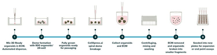
Figure 1. Workflow for automated CRC organoid culture on the CellXpress.ai system, showing steps from initial seeding in ECM, dome formation, growth monitoring, AI-triggered passaging, to expansion or assay setup
Plating into 96-well or 384-well plates
For testing compound activities in endpoint assays, organoids were expanded and then collected using a passaging protocol interrupted after the harvesting step or before the final dome plating step. Cell suspensions were used for seeding domes into the 96-well or 384-well plates. The suspension was seeded into 96-well plates (U-bottom Corning 4520) in 50% Matrigel domes, 15 μL per well (~200 organoids), using the 96-seeding protocol (Figure 9). After seeding, organoids were further processed for endpoint assays and imaged on the ImageXpress® HCS.ai High-Content Screening System.
3D colorectal cancer organoids (CRCs) expansion on the CellXpress.ai system (weeks 0–2)
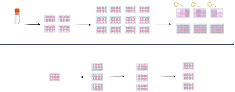
Figure 2. Workflow for automated CRC organoid culture on the CellXpress.ai system, showing steps from initial seeding in ECM, dome formation, growth monitoring, AI-triggered passaging, to expansion or assay setup
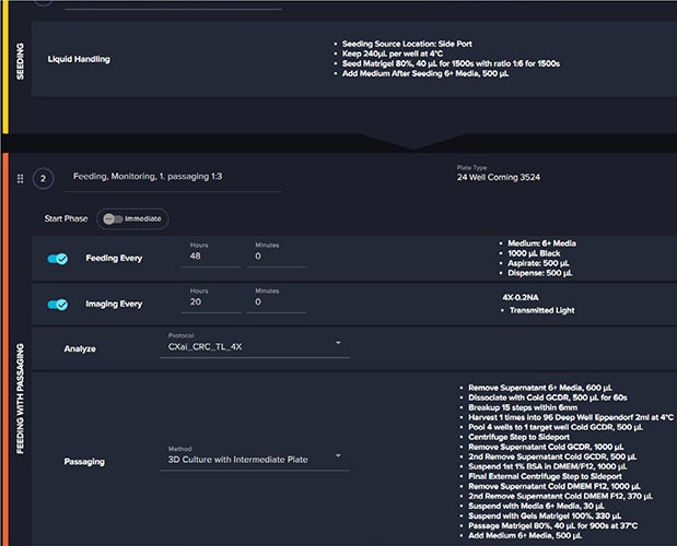
Figure 3. The picture illustrates the integrated workflow for seeding, feeding, imaging, and passaging organoids. Media exchanges and imaging are scheduled periodically, while passaging is triggered either manually or automatically based on AI-driven image analysis of organoid morphology and density. This protocol ensures consistent growth, reduces variability, and supports long-term maintenance.
Results
Automated expansion of patient-derived CRC organoids on the CellXpress.ai system
The CellXpress.ai system enabled fully automated seeding, feeding, imaging, and AI-guided passaging of patient-derived CRC organoids, expanding cultures from a single vial 3-fold, yielding mature organoids suitable for downstream assays. Automated seeding, scheduled media exchanges, and transmitted light imaging created a stable and reproducible environment for long-term culture (Figure 1).
Real-time imaging and IN Carta machine learning analysis provided continuous, non-invasive monitoring of organoid morphology and phenotypic state (Figure 3). Organoids were classified as young or old based on size, optical density, internal texture, and structural complexity, enabling objective quantification of culture maturity over time. During early culture (Day 0–1), organoids recovered from thawing or fragmentation and began progressive growth and lumen formation. With each 48-hour feeding cycle and daily imaging, the proportion of old organoids steadily increased until user-defined criteria for passaging were met.
AI-driven monitoring and passaging
Passaging was triggered either manually or automatically based on quantitative image analysis. A passaging event was initiated once the threshold of ≥50% old organoids in ≥80% of wells was reached. When triggered, the CellXpress.ai system executed automated dome breakage, pooling, centrifugation, mechanical fragmentation, and reseeding into fresh Matrigel domes (Figure 2). Cultures were expanded from one initial vial into twelve plates. These organoids were further maintained in culture in 1: 3 passaging for > 4 weeks. Imaging and analysis verified consistent organoid viability, morphology, and recovery following each passage cycle.
Harvest and seeding into 96-well plates
Expanded organoids were harvested using the same automated passaging workflow, with the process termination/ pause after fragment collection to allow reseeding into 96 or 384-well assay plates (Figure 5). Each 24-well plate supplied material for approximately three 96-well plates, with domes plated at 15 µL, at a density of ~200 organoids/well, ensuring uniform assay inputs. Confocal imaging confirmed structural integrity and epithelial organization of expanded organoids, with phalloidin highlighting actin architecture, ZO-1 labeling tight junctions, and DAPI marking nuclei (Figure 5). These structural markers validated that automated expansion preserved tissue architecture and phenotype required for downstream functional assays.
Together, these results demonstrate that the CellXpress.ai system effectively supports automated long-term maintenance, expansion, and generation of assay-ready organoids with minimal hands-on intervention.
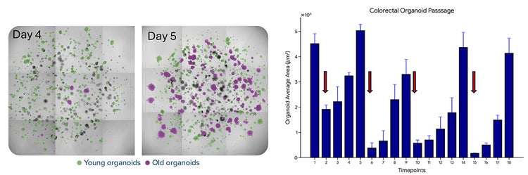
Figure 4. Organoid growth dynamics and passaging events during automated culture. Left: Brightfield images from Day 4 and Day 5 with AI segmentation masks showing young organoids (green) and old organoids (magenta). Right: Growth curve of average organoid area over time; dips in the curve correspond to passaging events, followed by organoid growth, demonstrating consistent recovery and expansion.
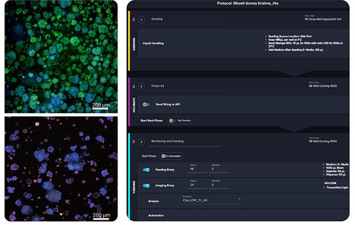
Figure 5. Expanded CRC organoids were seeded into 96-well plates for high-throughput assays. Left: Automated protocol for seeding, feeding, and monitoring on the CellXpress.ai system. Right: organoids were stained with AF488-labeled Phalloidin, showing actin cytoskeleton (green), fluorescently labeled antibodies against ZO-1 (red), and DAPI. Organoids were imaged using the HCS.ai confocal system. Organoids stained with AF488 phalloidin (green) and nuclei with DAPI (blue) confirm structural integrity, while ZO-1 (red) highlights tight junctions, validating epithelial organization.
Manual vs. automated workflow efficiency
All time estimates presented below assume the use of pre-optimized and fully configured protocols. Initial protocol optimization, method development, and troubleshooting require additional upfront time and are not included in these estimates.
CRC organoid culture: Manual vs. Automation
Maintaining CRC organoids using the Dome culture workflow is substantially more complex than conventional 2D cell cultures. Here is an estimate of hands-on labor required for manual CRC organoid culture compared with automated workflows using the CellXpress.ai system. The estimate is for the maintenance of twelve, 24-well CRC organoid plates during routine expansion and maintenance.
Manual CRC organoid culture: Breakdown
Manual maintenance of CRC organoids involves seeding of domes with organoids, careful media exchanges, frequent incubator-to-microscope transfers, and laborintensive passaging steps. Unlike 2D cultures, CRC organoid passaging requires gentle dissociation, cellular reagent matrix breaking, extensive mechanical dissociation, multiple cold centrifugation washes, and precise resuspension into fresh Matrigel. These steps demand sustained attention at the bench and contribute significantly to user fatigue due to repetitive steps, pipetting, and variability.
Automation with the CellXpress.ai system: Walkaway
The CellXpress.ai system automates critical CRC organoid culture steps, including media exchange, imaging, morphological analysis, and ECM dome breakage for passaging. Once workflows are configured, manual interaction is limited to reagent replenishment, consumable loading, and protocol review. Weekly hands-on time is reduced to approximately one hour, with most culture operations occurring unattended within a controlled environment.
This shift converts repetitive, high-risk manual labor into reproducible, traceable workflows. As the experiment scale increases, time savings become exponential, making the CellXpress.ai system a critical platform for complex 3D culture.
(media changes)
(3D CRC organoids)
Summary
The CellXpress.ai system enables robust, reproducible, and scalable expansion of patient derived CRC organoids by integrating automated liquid handling, environmental control, and AI-driven image analysis into a unified culture system. Starting from a single vial, the platform expanded CRC organoids 3-fold with consistent morphology and long-term viability. Automated seeding, scheduled feeding, and daily transmitted light imaging maintained stable growth, while the IN Carta software’s deep learning segmentation and Phenoglyphs classification provided unbiased assessments of organoid maturity to guide passaging decisions. Expanded organoids were then harvested and transitioned into high-throughput assay formats, with confocal imaging confirming intact epithelial architecture, including actin cytoskeleton and tight junctions.
In addition to improving reproducibility, the CellXpress. ai system substantially reduces the hands-on burden associated with CRC organoid culture. Routine operations that typically require extensive manual effort are consolidated into automated workflows, allowing scientists to focus on experimental design and downstream analysis rather than repetitive culture maintenance. This combination of biological consistency and operational efficiency supports reliable production of assay-ready organoids for translational CRC research. This level of automation brings practical convenience to organoid workflows, allowing scientists to shift time and attention from routine maintenance toward higher value experimental decisions.
Conclusion
The CellXpress.ai system transforms CRC organoid culture by pairing automation with AI-enabled quality control to deliver consistent, scalable expansion and reliable longterm maintenance of patient derived organoids. The platform standardizes seeding, feeding, imaging, and passaging, minimizing variability and ensuring high-quality organoids suitable for functional screening and imagingbased assays. Importantly, by automating the most laborintensive steps of organoid culture, the CellXpress.ai system reduces routine hands-on time to a fraction of what is required manually, providing meaningful time savings without compromising biological performance. This integrated, data-driven approach accelerates organoidbased discovery workflows and strengthens the use of 3D cancer models in translational research.
HUB Organoid Technology used herein was used under license from HUB Organoids. To use HUB Organoid Technology for commercial purposes, please contact bd@huborganoids.nl for a commercial use license.